Enzymes

Enzymes, proteins, riboproteins and signaling pathways.

Enzymes

Enzymes are organic catalysts, chemicals that increase the rate at which reaction equilibrium is achieved without themselves being permanently changed by the reaction.

Endothermic reactions, such as the steps in biosynthesis of macromolecules, require heat input, while exothermic reactions release heat. Thermochemistry deals with the heat exchange of chemical reactions, while reaction energetics deals with the dynamics of chemical reactions.

Although enzymes alter k, the rate of a reaction, they do not alter Keq, the actual equilibrium point. However, if the products of a reaction are removed by a second reaction, then the product side of the reaction equation will be favored because equilibrium tends toward maintaining the ratio of products to reactants.

Constitutive enzymes are always produced at roughly the same concentration regardless of the composition of the medium (glycolysis, TCA cycle). Conversely, inducible enzymes are produced in response to a particular substrate, being produced only when needed. In induction, the inducer substrate, or a structurally similar compound, promotes formation of the enzyme. Conversely, the production of repressible enzymes downregulated by environmental conditions, such as the presence of the end product (corepressor) of a pathway in which the enzyme normally participates.

Regulation of enzymatic function:
Enzymes often function in metabolic chains in which regulation occurs by specific feedback mechanisms. Individual metabolic reactions are typically managed in the forward and reverse directions by structurally distinct enzymes, which permits regulation of metabolic pathways.

In bacterial cells, regulation of enzymatic reactions proceeds by:
1) control or regulation of enzyme activity by feedback inhibition or end product inhibition, which regulate biosynthetic pathways, or
2) control or regulation of synthesis of inducible or repressible enzymes, by
a) negative control as end-product repression, which downregulates enzyme synthesis and associated biosynthetic pathways,
b) positive control as enzyme induction and catabolite repression, which upregulate enzyme synthesis and associated degradative pathways

Metabolic regulation is often implemented through allosteric enzymes, which possess, as do allosteric proteins, multiple shape-changing subunits with distinct active sites. Allosteric enzymes change shape between active and inactive forms in response to the binding of substrates at the active site, or to binding of regulatory molecules at other sites. Because the reaction rates of allosteric enzymes can be regulated by only small changes in substrate concentration, allosteric enzymes are employed by cells to regulate metabolic pathways in which the concentration of cellular substrates fluctuates over narrow concentration ranges. In the simplest case in which an allosteric enzyme with a positive effector site has an active and an inactive form, the alteration in reaction rate in response to increasing substrate concentration typically displays an "S-shaped" curve. After binding of a molecule to the positive effector site of an allosteric enzyme, the second and subsequent substrates bind readily because binding of the effector molecule has induced a favorable structural change. This response is termed "cooperativity," and the S-shaped curve indicates the cooperative binding of substrate. Conversely, for allosteric enzymes with negative effector sites, binding to the allosteric site inactivates further substrate binding to the active site.

MIT Biology Hypertextbook: Enzyme Mechanisms: "Not all proteins are enzymes, but most enzymes are proteins (the exception is catalytic RNA). [Enzymes are catalysts employed in cellular reactions.] A catalyst is a molecule which increases the rate of a reaction but is not the substrate or product of that reaction. A substrate (A) is a molecule upon which an enzyme acts to yield a product (B).

A –––→ BEnz

The free energy of this reaction is not changed by the presence of the enzyme, but, for a favored reaction (where ΔG is negative), the enzyme can speed it up."

Enzymes couple with substrates in transitional states, effecting conformational changes (3D structure) that facilitates transition to products.

The classification and naming of enzymes, according to the EC, depends upon their function in the reactions that they catalyze:
1. EC1. Oxidoreductases alter the oxidation state of molecules by transfer of electrons (often as hydride ions H−).
2. EC 2. Transferases transfer chemical groups from one molecule to another (not to be confused with cofactors which carry groups).
3. EC 5. Isomerases transfer chemical groups within molecules.
4. EC 3. Hydrolases add or remove H2O from molecules.
5. EC 4. Lyases manipulate double bonds in elimination reactions.
6. EC 6. Ligases condense bonds between C- and S/N/C/O, using energy from ATP

Similarly, enzymes such as RNA polymerase are named for their actions, where RNA polymerase is a common name for ATP:[DNA-directed RNA polymerase] phosphotransferase.

Specific enzymes:
DNA polymerases & reverse transcriptase : base excision repair = DNA glycosylase & AP endonuclease & Fen1 protein : hOGG1 DNA repair : helicases : RNA polymerase : ribozymes : ribozymes in repair of RNA and DNA :

Alphabetical: Enzymes →reaction→ A: ~ adenylate cyclases : AP endonuclease (Ape1) : aspartate aminotransferase ~ ATPases ~ : B: base excision repair : C: ~ cyclin-dependent kinases ~ cytochrome c oxidase s: D: DNA glycosylase : DNA Ligase I : DNA polymerases : DNA polymerase I : DNA polymerase beta : DNase IV : E: exonuclease 1 : exosome : F: Fen1 : Flap Endonuclease FEN-1 : G: general transcription factors : glucose-6-phosphate dehydrogenase : →glutamate-dehydrogenase→ : H: hOGG1 : hOGG1 oxoG oxoG repair : L: LigIII : M: MAP kinase : Msh2-Msh3 : MutS, MutL, and MutH : N: NADH dehydrogenase : nucleotide excision repair : O: 8-oxoguanine glycosylase : oxoG oxoG repair hOGG1 : P: PCNA : ~ phosphatases ~ phospholipases ~ phosphodiesterases ~ phosphorylases ~ 6-phosphogluconate dehydrogenase ~ protein kinases ~ pyruvate carboxylasepyruvate carboxylase→: pyruvate dehydrogenasepyruvate dehydrogenase reaction : R : ~ receptor tyrosine kinases ~ RNA polymerase : replication : Replication factor C : reverse transcriptase : ribozymes : ribozymes in repair of RNA and DNA : ribulose bisphosphate carboxylase/oxygenase : RNA polymerase II : Rubisco : S: ~ serine/threonine kinases ~ spliceosomal-mediated RNA trans-splicing SMaRT : succinate dehydrogenase : T: transaldolase : transketolase : trans-splicing ribozymes : U: UvrD : X: XRCC1 :

MIT Biology Hypertextbook on Enzyme Biochemistry : Chemical Energetics : Enzyme Mechanisms : Enzyme Kinetics : Feedback Inhibition

| 0 Guide-Glossary

reaction energetics

Reaction energetics describes the energetic principles that underlie chemical reactions, phase changes, and solution formation. In essence, thermodynamically favorable reactions move readily from reactants to products, and are accelerated in the presence of specific catalysts or enzymes.

For a reaction A + B → C + D
A and B are the reactants and C and D are the products. Energetically favorable reactions proceed spontaneously from reactants to products. In such a case the energy state of reactants is lower than the energy state of products, meaning that the reaction proceeds in a downhill direction in terms of chemical energy. However, to reach this downhill state, the reactants must pass over a 'speed bump', an energetic barrier.

Catalysts lower the energetic barrier between substrates and products yet catalysts emerge unchanged from the reaction. Biological catalysts are called enzymes – these are usually proteins, but may be molecules of RNA (ribozymes).

Although enzymes alter k, the rate of a reaction, they do not alter Keq, the actual equilibrium point. However, if the products of a reaction are removed by a second reaction, then the product side of the reaction equation will be favored.

Enzymes increase the rate of reactions by virtue of a transient binding of substrate (A and/or B) to the active site of the enzyme. This binding of substrate to enzyme occurs at the active site where it is stabilized by numerous weak interactions (hydrogen bonds, electrostatic interactions, hydrophobic contacts, and van der Waals forces). The enzyme-substrate complex dissociates into enzyme (in original state) and products (C,D).

Endothermic reactions, such as the steps in biosynthesis of macromolecules, require heat input, while exothermic reactions release heat.

| 0 Guide-Glossary

bonds

Atoms interact and some atoms form relatively stable chemical bonds with other atoms.

Weakest to Strongest :
intermolecular van der Waals electrostatic interaction :
intermolecular hydrogen bond :
intramolecular covalent bond :
intramolecular ionic bond :

Atomic orbitals are often represented as electrons spinning around the nucleus (top). However, orbital shells actually represent the volume in which the wave-form electron is most likely to be located (bottom). As such, orbitals are more akin to a cloud around the nucleus. The location of electrons within orbitals is described mathematically by the Schrödinger equations. Computer simulations reveal probability distributions for orbitals. Table of images of orbitals / Scatter plot of probabilities / interactive / virtual text / virtual text orbitals / download audio-anim of H2 bonding / audio-anim of hybridization of s p orbitals / voxel movie of orbital / movie 2 / movie 3 / movie 4 /

Ionic bonds are strong bonds formed when one atom is sufficiently electronegative to remove an electron from a sufficiently electropositive atom – creating negative and positive ions that are attracted by virtue of their opposite polarity.

When electrons are shared between two atoms of the same element, the electrons are shared equally, creating a non-polar covalent bond. When electrons are shared between atoms of different elements, the electrons are not shared equally, resulting in a polar covalent bond in which the increased-probability cloud over one atom has a slightly negative charge compared to a slightly positive charge over the other – a dipole (δ+, δ-).

Hydrogen bonds are intermolecular attractions between a hydrogen atom and a small, electronegative atom in a neighboring atom share a dipole-dipole attraction (δ+ δ-). Although stronger than most other intermolecular forces, a hydrogen bond is much weaker than either an ionic bond or a covalent bond. Within macromolecules such as proteins and nucleic acids, hydrogen bonds can exist between two parts of the same molecule and constrain the molecule's 3D shape.

| 0 Guide-Glossary

adenylate cyclases

Adenylyl (adenylate) cyclases are enzymes, which cross the membrane twelve times (right) and which convert ATP to the second-messenger cAMP (3',5' cyclic AMP) and pyrophosphate (below left). Likewise, guanylate cyclases convert GTP to the second messenger, cGMP. Adenylyl cyclases are coincidence detectors, meaning that they are only activated by several different signals occurring together – they are modulated by G-proteins, forskolin, Ca2+/calmodulin, and other class-specific substrates.

| 0 Guide-Glossary

AID

Activation-induced (cytidine) deaminase (AID) is a 24 kDa enzyme currently considered the master regulator of secondary antibody diversification because it is involved in the initiation of three distinct immunoglobulin diversification processes: somatic hypermutation (SHM), class-switch recombination (CSR), and gene-conversion (GC).

AID-generated somatic hypermutations affect the variable (V) regions of genes encoding immunoglobulins. SHM is restricted to VDJ regions and their adjacent flanks in immunoglobulin (Ig) genes, whereas constant regions are spared because AID does not gain access to the 5' and constant regions of Ig genes.[r1] Mutations occur after about 100 nucleotides downstream of the promoter and extend to 1-2 kb. Somatic (hyper)mutation affects only individual cells of the immune system, so the programmed mutations that it generates are transmitted only within the particular cell line (somatic) and are not transmitted to the organism's offspring.

Following activation of naïve B cells and during the subsequent antigen-stimulated proliferation of B cells, the gene locus for the Ig-BCR experiences a highly accelerated rate of somatic mutation (increased by a factor of 10^5 to 10^6). This acceleration is attributable to the enzyme activation-induced (cytidine) deaminase (AID), which extracts the amino group from a deoxycytidine base in DNA, converting deoxycytidine to deoxyuracil. Deoxycytidine is a nucleoside formed through attachment of the nucleobase cytosine to a deoxyribose ring via a β-N1-glycosidic bond, and deamination of cytosine generates uracil in deoxyuridine (dUMP) []im C to U[].

AID-catalyzed deamination of deoxycytidine creates a single nucleotide polymorphism (SNP) in the DNA strand by generating a uracil:guanine mismatch. The nucleobases that normally occur in DNA are adenine paired with thymine, and cytosine paired with guanine. Uracil is normally found only in RNA, where it is paired with adenine.

A high-fidelity base excision repair enzyme, uracil-DNA glycosylase (UNG2), excises the alien uracil nucleobase, then error-prone DNA polymerases complete the base-excision repair. During this base-excision repair, incorrect nucleobases may be substituted at or adjacent to the original C to U mutation site. Mispairing (transition) mutations are susceptible to indels - insertions and deletions. (Such mutation vulnerable areas in the genome are termed 'hotspots', and they have played a significant role in biological evolution.)

Thus, while AID deamination generates mutagenic U:G mismatches, DNA-incorporated dUMP generates U:A pairs that are not directly mutagenic, but which may be cytotoxic. Usually, deleterious mutations that could result from uracil withiin DNA are prevented by error-free base excision repair.

However, B-cells employ uracil in DNA as a physiological intermediate in the somatic hypermutation processes that promote secondary antibody diversification in adaptive immunity. Here, activation-induced cytosine deaminase (AID) introduces template uracils that provide for GC to AT transition mutations at the Ig locus after replication. When the base excision repair enzyme uracil-DNA glycosylase (UNG2) excises uracil, error-prone DNA polymerases may causes other mutations at/near the abasic site of the Ig locus.

Together, these processes are central to the somatic hypermutation (SHM) mechanism that increases immunoglobulin diversity. Similarly, AID and UNG2 are also essential for the generation of strand breaks that initiate the process of class-switch recombination (CSR).

[r1] The very 5' end and the constant region of Ig genes are spared from somatic mutation because AID does not access these regions. Longerich S, Tanaka A, Bozek G, Nicolae D, Storb U. J Exp Med. 2005 Nov 21;202(10):1443-54. [Free Full Text Article]

| 0 Guide-Glossary

Akt

Akt1 is 'thymoma viral proto-oncogene 1', a synonym for protein kinase B, and is a serine/threonine kinase that promotes cellular survival.

: 14-3-3 : activation of Akt1 : Akt1 action : Bad : Bad/Bcl-XL : Bcl-XL : c-Akt : caspase 9 : cell survival pathways : forkhead : growth factors : hTERT : IkB kinases : IKKα : ILK : integrin : NF-κβ : surface receptors : PDK1 : PI3K : PIP control : PTEN : v-akt : Wortmannin :

Specific protein kinases transfer a phosphate group from a donor such as ATP to amino acid acceptors in proteins, while protein phosphatases remove the phosphate groups that have been attached by protein kinases. Proto-oncogenes participate in a variety of normal cellular functions, but have the potential to tranform into cellular oncogenes when mutated. Proto-oncogenes normally function in the various signal transduction cascades that regulate cell growth, proliferation and differentiation. Cellular proto-oncogenes resident in transforming retroviruses are designated as c- (cellular origin) as opposed to v- (retroviral origin).

Akt1 activation requires PDK1 phosphorylation of Thr308 in the activation domain and is dependent on the products of phosphatidylinositol (PI) 3-kinase (PI3K), phosphatidylinositol 3,4 bisphosphate (PIP2) and phosphatidylinositol 3,4,5 trisphosphate (PIP3). When activated, Akt exerts anti-apoptosis effects through phosphorylation of substrates that directly regulate the apoptosis machinery (Bad or caspase 9), or phosphorylation of substrates that indirectly inhibit apoptosis (human telomerase reverse transcriptase subunit (hTERT), forkhead transcription family members, or IkB kinases). Akt promotes survival in vitro when cells are exposed to different apoptotic stimuli such as growth factor deprivation, UV irradiation, matrix detachment, cell cycle discordance, DNA damage, and administration of anti-Fas antibody, TGF-β, glutamate, or bile acids.

Akt is the cellular homologue of the product of the v-akt oncogene and has 3 isoforms, Akt1, 2, and 3 (or PKB-α, -β, and -γ). Akt is activated by many growth factors, including IGF-I, EGF, βFGF, insulin, interleukin-3, interleukin-6, heregulin, and VEGF. Full activity of all three isoforms requires phosphorylation of both a site in the activation domain and another site in the C-terminal hydrophobic motif.

Many cell surface receptors induce second messengers that activate phosphatidylinositide-3-OH kinase (PI3K). Because Akt is located downstream of PI3K Akt functions as part of a wortmannin-sensitive signaling pathway. PI3K generates phosphorylated phosphatidylinositides in the cell membrane, which bind to the amino-terminal pleckstrin homology (PH) domain of Akt. The phosphatidylinositides, PI-3,4-P2 and PI-3,4,5-P3 also activate phosphoinositide-dependent kinase-1 (PDK1) which phosphorylates Thr308 of membrane-bound Akt. The Ser473 is phosphorylated by integrin-linked kinase (ILK).

Activated Akt promotes cell survival via two distinct pathways:
1) Akt inhibits apoptosis by phosphorylating the Bad component in the Bad/Bcl-XL complex. When phosphorylated, Bad binds to protein 14-3-3, leading to dissociation of the Bad/Bcl-XL complex and permitting cell survival.
2) Alternatively, Akt activates IKK-α, which ultimately leads to NF-κβ activation and cell survival.

Cellular levels of PIP2 and PIP3 are controlled by the tumor suppressor, dual-phosphatase PTEN, which dephosphorylates PIP2 and PIP3 at the 3' position.[2]

Wortmannin is a fungal metabolite that is a specific inhibitor of PI3Ks, though it can also inhibit mTOR, DNA-PK, some phosphatidylinositol 4-kinases, myosin light chain kinase (MLCK) and mitogen-activated protein kinase (MAPK).

: 14-3-3 : activation of Akt1 : Akt1 actionapoptosis : Bad : Bad/Bcl-XL : Bcl-XLBcl-2 : c-Akt : caspase 9caspases : cell survival pathwayscellular survivaldeath receptor : forkhead : growth factors ~ growth factors : hTERT : IkB kinases : IKKα : ILK : integrin ~ integrins : NF-κβ : surface receptorsreceptor proteins : PDK1PDK1 : PI3KPI3K : PIP control : PTENPTENreceptor proteins ~ second messengerssignal transduction : v-akt : Wortmannin :

Top

| 0 Guide-Glossary

ATPases

ATP synthase is the final enzyme of oxidative phosporylation. It is located in the inner mitochondrial membrane and utilizes the proton-gradient potential energy generated by an electron transfer chain to phosphorylate ADP to ATP.

The F1Fo ATP synthase is the commonest chemi-mechanical motor found in nature. The Fo motor is an integral energy membrane protein complex that propels converts transmembrane chemical gradients into the rotary mechanical motion of the γ-subunit during ATP synthesis. The F1 motor is a peripheral membrane protein complex connected to the Fo motor. The F1 motor employs the Fo-propelled rotation to drive the sequential condensation of ADP with Pi at its three catalytic sites. ATP syntase can also be run in reverse as the chemical energy of ATP is employed to generate mechanical motion or to pump protons against a chemical potential.

The ATPase-driven rotation of the γ-subunit utilizes sequential conformational changes of each of three catalytic sites on the β-subunits. Because there are three catalytic sites on the enzyme, each ATPase event induces a 120 degree rotation of the γ-subunit. The conformational changes facilitate ATP synthesis by altering the dissociation constant of ATP relative to ADP. The conformations of the β-subunits are staggered such that all three conformations are present at any particular moment. Structural asymmetry of the catalytic sites plus their differences in affinity for ATP occur only when the nucleotide is bound as a complex with the Mg2+ cofactor, indicating that ATP synthesis depends on changes in metal ligands. Calcium, which can bind to two more ligands than can Mg2+, is an effective cofactor of the ATPase reaction. However, Ca2+ activity does not pump a transmembrane proton gradient, implying that Ca2+ couples with the protein at positions that couple ATP hydrolysis to the generation of rotational torque on the γ-subunit.

| 0 Guide-Glossary

cAMP-dependent protein kinase

cAMP-dependent protein kinase (PKA) is a serine/threonine kinase with catalytic (protein phosphorylating) activity that is modulated by cAMP levels. Specific protein kinases transfer a phosphate group from a donor such as ATP to amino acid acceptors in proteins, while protein phosphatases remove the phosphate groups that have been attached by protein kinases.

PKA is highly conserved with receptor tyrosine kinases.

| 0 Guide-Glossary

cyclin-dependent kinases

Cyclin-dependent kinases (CDKs) play a role in regulation of transcription and in mRNA processing, and in regulation of the cell cycle. Specific protein kinases transfer a phosphate group from a donor such as ATP to amino acid acceptors in proteins, while protein phosphatases remove the phosphate groups that have been attached by protein kinases.

The activity of cyclin-dependent kinases is modulated by binding to cyclins, which are proteins so-named because their levels vary periodically during the cell cycle. The binding of cyclins to cyclin-dependent kinases regulates CDK activity, selecting the proteins to be phosphorylated. Although levels of CDK-molecules are constant during the cell cycle, their activities vary because of the regulatory function of the cyclins. Thus, periodic protein degradation is an important general control mechanism of the cell cycle.

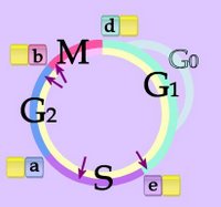
Right - Cell Cycle - click to enlarge image. Phases G1 and G0, S, G2, M. Checkpoints - purple arrows at G1-S transition, during S phase, G2-M transition, anaphase of mitosis. CDKs (yellow) are modulated by association with a series of fluctuating cyclins (d, e, a, b.) The cycle is divided into non-mitotic interphase (beige) with G1 or G0, S, and G2 phases followed by mitosis (pink).

Cyclins were conserved during evolution. Around ten different cyclins have been found in humans. CDK and cyclin together drive the cell from one cell cycle phase to the next. Levels of cyclins D (G1), E and A (S), and B and A (mitotis, M) fluctuate during the cell cycle (d,e,a,b) , and binding of appropriate cyclins to the cyclin-dependent kinases (CDKs) stimulating phosphorylation and activation.

CDKs are an example of enzymes that possess separate phosphorylation sites for the activation or inhibition of functional regulation. CDKs are a subclass of serine/threonine protein kinases that can be either activated or deactivated depending upon the specific amino acid residue that is undergoing phosphorylation.

Studies with yeast and embryonic cells suggest that mitosis is triggered by the periodic activation of cdc2 kinase (cdk1), named for its role as a CDC-gene (cell division cycle gene). This member of the Ser/Thr protein kinase family is a catalytic subunit of the highly conserved protein kinase complex known as M-phase promoting factor (MPF), which is essential for G1/S and G2/M phase transitions of eukaryotic cell cycle. Mitotic cyclins form stable associations with cdk1 (cdc2), and function as regulatory subunits. Because the kinase activity of cdk1 is controlled by cyclin accumulation and destruction duringthe cell cycle, phosphorylation and dephosphorylation of cdk1 play important regulatory roles in cell cycle control. More than one hundred cell division cycle genes (CDC genes) are specifically involved in cell cycle control

Top

| 0 Guide-Glossary

ceruloplasmin

Ceruloplasmin, (Cp) caeruloplasmin, ferroxidase, or iron(II):oxygen oxidoreductase is an acute phase protein synthesized in response to pro-inflammatory cytokines in the inflammatory response (acute and chronic).

Ceruloplasmin (ferroxidase) is an enzyme that catalyzes oxidation of ferrous iron (Fe2+) to ferric iron (Fe3+), rendering iron suitable for binding and plasma transportation by transferrin.
Cp is also reported to oxidize LDL, so, since oxidized LDL (Ox-LDL) is a well-known atherogenic factor, elevated serum Cp is expected to act as an atherogenic factor. Elevated level of Ox-LDL inhibits nitric oxide (NO) production, and a decreased level of NO impairs the endothelium-dependent relaxation of arteries, providing a factor causing atherosclerosis.

Ninety percent or more of total serum copper is located in ceruloplasmin, though albumin is the chief transport protein for copper. Levels of ceruloplasmin are elevated in acute and chronic inflammation, rheumatoid arthritis, lymphoma, carcinomas, leukemia's, Hodgkin disease, primary biliary cirrhosis, systemic lupus erythematosus, pregnancy. Levels are depressed in copper deficiency, Vitamin C overdose, excessive therapeutic zinc, and rare disorders of copper metabolism and/or storage (aceruloplasminemia, Menke's kinky hair syndrome, Wilson's disease (hepatoenticular degeneration)). Aceruloplasminemia is caused by mutations in the gene encoding ceruloplasmin on chromosome 3q (bands 3q23-q25).

tags

| 0 Guide-Glossary

Fyn

The FYN gene that encodes the enzyme Fyn is a member of the protein tyrosine kinase oncogene family and is related to SRC, FGR, and YES.

Fyn is a membrane-associated tyrosine kinase implicated in the control of cell growth. Fyn protein associates with the p85 subunit of phosphatidylinositol 3-kinase and interacts with the fyn-binding protein. There exist alternatively spliced transcript variants encoding distinct isoforms of Fyn.

The intracellular tails of CD3 molecules contain a single conserved motif termed the immunoreceptor tyrosine-based activation motif (ITAM) that is essential for TCR signaling. Phosphorylation of CD3's ITAM enables the CD3 chain to bind Fyn, making this kinase important in the T cell's signaling cascade.

| 0 Guide-Glossary

GTPases

GTPases hydrolyze GTP to GDP + Pi in the highly conserved domains of G-proteins, which are associated with GPCRs (guanine nucleotide-binding protein-coupled receptors, G-protein coupled receptors, serpentine receptors, 7TM receptors, or heptahelical receptors).

G-proteins, guanine nucleotide binding proteins, are membrane-associated trimeric proteins that associate with receptors to participate in cellular signaling. Large G-proteins comprise the GDP/GTP-binding α-subunit, which is asssociated with the βγ-subunits. Small monomeric G-proteins like Ras are important molecular switches that also participate in signal transduction pathways.

_______________GDP________________________ GTP

In association with a GPCR, ligand-binding by the transmembrane receptor induces the α-subunit to exchange its bound GDP molecule for a GTP molecule, upon which the G-protein dissociates from the βγ-subunit and the receptor. The receptor is now free to engage another G-protein trimer, and both the α-GTP- and βγ-subunits are free to activate signaling cascades (second messenger pathways), gate ion channels, and activate effector proteins. As a GTPase, the α-GTP-subunit hydrolyzes its attached GTP to GDP, freeing the α-subunit to re-associate with the βγ-subunit and the receptor, initiating a new cycle. Thus, the G-protein acts as both an amplifier and a transducer of the signal.

GTPase superfamily functions:
a. Signal transduction at the intracellular domain of transmembrane receptors (GPCRs), including sensory perception (taste, smell, light).
b. Protein assembly (translation) at ribosomes.
c. Regulation of cell cycle, and cellular differentiation.
d. Active transport and transmembrane protein transport (translocation).
e. Transport of vesicles within the cell, where GTPases control the assembly of vesicle coats.

| 0 Guide-Glossary

IκB kinase

Activation of IκB kinase (IKK) by stress signals stimulates phosphorylation of two serine residues in the regulatory domain 0f Inhibitor of kappa B (IκB), targetting the IκB molecules for ubiquitin/proteasome degradation, and releasing NF-κB from inhibition. (Intact Inhibitor of kappa B (IκB) inactivates NF-κB by sequestering NF-κB dimers within the cytoplasm.) Physiological activities mediated by NF-κB include cellular proliferation, and inflammatory, immune, and cellular survival responses.

| 0 Guide-Glossary

MAPKs

Mitogen activated protein kinases (MAP kinases, MAPKs) are serine/threonine kinases that are activated by mitogens, and act as switch kinases that convert information of increased intracellular tyrosine phosphorylation into serine/threonine phosphorylation. Specific protein kinases transfer a phosphate group from a donor such as ATP to amino acid acceptors in proteins, while protein phosphatases remove the phosphate groups that have been attached by protein kinases. Maximal MAP kinase activity requires phosphorylation of both tyrosine and threonine residues.

The MAPK signaling cascade is:mitogen → MAPKK kinase (MAPKKK) → MAPK kinase (MAPKK) → MAP kinase (MAPK) → signaling

All eukaryotic cells possess multiple MAPK pathways [K], which coordinately regulate diverse cellular activities running the gamut from gene expression, mitosis, and metabolic regulation to motility, survival and apoptosis, and cellular differentiation.

To date, five distinct groups of MAPKs have been characterized in mammals: extracellular signal-regulated kinases (ERKs) 1 and 2 (ERK1/2), c-Jun amino-terminal kinases 1, 2, and 3 (JNKs, or SAPK, stress-activated protein kinases), p38 isoforms α, β, γ, and δ, ERKs 3 and 4, and ERK5 (reviewed in references 25 and 103).[s-fft]

Other MAP kinases include: microtubule associated protein-2 kinase (MAP-2 kinase), myelin basic protein kinase (MBP kinase), ribosomal S6 protein kinase (RSK-kinase) and EGF receptor threonine kinase (ERT kinase). MAP kinases include extracellular-signal regulated kinases (ERKs), with activators that include mitogens: Ras [fft], polypeptide growth factors PDGF, CSF-1, IGF-1, EGF, insulin, and PMA.

Top

| 0 Guide-Glossary

MEK

MEKs are dual-specifity threonine and tyrosine recognition kinases that activate extracellular signal-regulated kinase (ERK) isoforms of mitogen-activated protein kinases (ERK-MAPKs). MEKs are, in turn, activated by phosphorylation.

MEK1 acts as a convergence point for the multiple protein kinases involved in MAPK activation[1] Specific protein kinases transfer a phosphate group from a donor such as ATP to amino acid acceptors in proteins, while protein phosphatases remove the phosphate groups that have been attached by protein kinases.

MEK act as substrates for several protein kinases including the Rafs (c-, A- and B-), Mos, Tpl-2, and MEKK1. Raf and MEKK mainly phosphorylate MEKs at serine residues (218 and 222 in rat MEK1). Raf and MAPK/extracellular signal-regulated kinase kinase (MEKK) independently phosphorylate and activate MEK-1, establishing a protein kinase signaling cascade whose activity is controlled by G-protein linked effectors and tyrosine kinases. Raf phosphorylation is reported to increases recombinant MEK1 activity two-fold, while MEKK phosphorylation of recombinant MEK1 increases MEK1 activity five-fold.

MAPK phosphorylates MEK1 at sites (threonine) different than those phosphorylated by Raf and MEKK (serine and tyrosine)[s4]. MEK1 acts as a convergence point for the multiple protein kinases involved in MAPK activation.[s7] MEKK and Raf regulate MEK-1 activity by phosphorylation of common residues and thus, two independent protein kinases converge at MEK-1 to regulate the activity of MAPK. Phosphorylation of MEK-1 by MAPK does not affect MEK-1 kinase activity. Constitutive activation of MEKs depends on introduction of acidic residues and truncation of an alpha-helical region in the N-terminal domain. Mutation of the serine residues to alanine generates dominant-negative proteins that have been used to determine requirement for the ERK pathway. The major site of MAPK phosphorylation in MEK-1 is threonine 292, and site-directed mutagenesis of threonine 292 to alanine eliminates 90% of MAPK catalyzed phosphorylation of MEK-1 yet does not inhibit MEK-1 autoactivation.

Two related genes encode MEK1 and MEK2 which differ in their binding to ERKs. MEKs do not phosphorylate either SAPK or p38 MAPK. [ut]

The Raf/mitogen-activated protein kinase (MAPK/ERK) kinase, (MEK)/mitogen-activated protein kinase (MAPK), and the phosphatidylinositide-3-OH kinase (PI3K)/3-phosphoinositide-dependent protein kinase-1 (PDK1)/Akt pathways play central roles in the regulation of cell survival and proliferation.

MEK-1 is a dual threonine and tyrosine recognition kinase that phosphorylates and activates mitogen-activated protein kinase (MAPK). MEK-1 is in turn activated by phosphorylation.[1]

| 0 Guide-Glossary

MPF

M-phase promoting factor is a complex of cyclins with the M-phase cyclin-dependent kinases, which initiates metaphase assembly of the mitotic spindle, breakdown of the nuclear envelope, and condensation of chromosomes.

| 0 Guide-Glossary

mTOR

mTOR is a serine/threonine kinase that regulates regulates translation and cell division, and is officially termed FRAP1 for FK506 binding protein 12-rapamycin associated protein 1.

mTOR stands for mammalian target of rapamycin, which is a fungal derivative that halts protein synthesis by complexing with immunophilin FK-506 binding protein FKBP12 peptide prolyl cis/trans isomerase. The mTOR protein kinase receives stimulatory signals from nutrients as well as Ras and phosphatidylinositol-3-OH kinase (PI3K) downstream from growth factors, so it functions as a signaling molecule and a critical growth-control node.

FRAP1 (mTOR) is an evolutionarily conserved member of the phosphoinositol kinase-related kinase (PIKK) family that includes DNA-PK, ATM, ATR and several other proteins. mTOR participates in the regulation of cell growth through initiation of gene translation in response to nutrients by integratating input from multiple upstream pathways, including growth factors, mitogens, leucine, insulin, and nutrients. mTOR initiates translation by activating the ribosomal p70S6k protein kinase (S6K1) and by inhibiting the eIF4E inhibitor 4E-BP1. FRAP1 is considered to be involved in numerous additional cellular functions including actin organization, membrane trafficking, secretion, protein degradation, protein kinase C signaling, ribosome biogenesis and tRNA synthesis. mTOR may contribute to the regulation of two pathways, referred to as TORC1 and TORC2 (for TOR Complex 1 and 2).

| 0 Guide-Glossary

PDK1

3-Phosphoinositide-dependent protein kinase-1 (PDK1) is a serine /threonine protein kinase that phosphorylates and activates PKB/AKT (Thr308 and Ser473), p70 S6 kinase, p90 ribosomal protein S6 kinase (RSK), PKA, PKC and serum and glucocorticoid-inducible kinase (SGK). (1, 2, 9)

Following stimulation by receptor tyrosine kinases, PI3K is activated and generates the phospholipid second messengers, PtdIns(3,4,5)P 3 and PtdIns(3,4)P 2. These second messengers then emplot diverse mechanisms to mediate the phosphorylation and activation of PDK1 targets (a-st).

Cellular stimulation with pervanadate and IGF-1 results in a significant increase in PDK1 activity and its translocation to the plasma membrane along with increased phosphorylation of tyrosine residues (pk). Phosphorylation on the activation loop at Ser241 alone is necessary for PDK1’s activity and a mutation of single nucleotide (SNP) abolishes its activity (cs).

The phosphatidylinositide-3-OH kinase (PI3K)/3-phosphoinositide-dependent protein kinase-1 (PDK1)/Akt and the Raf/mitogen-activated protein kinase (MAPK/ERK) kinase (MEK)/mitogen-activated protein kinase (MAPK) pathways play central roles in the regulation of cell survival and proliferation.

Pyruvate dehydrogenase kinase, isoenzyme 1 or 3-phosphoinositide-dependent kinase-1 (PDK1) contains an amino-terminal kinase domain and a carboxyl-terminal pleckstrin homology (PH) domain. PDK1 appears to be conserved throughout evolution (27-31). Although the PDK1 PH domain binds the lipid products of the phosphatidylinositol 3-kinase (PI3K) reaction, binding of these lipids does not alter PDK1 activity, rather it is necessary to localize PDK1 to the plasma membrane. Sphingosine, another biologically active lipid, activates PDK1 toward a variety of substrates (26). It is well established that PDK1 phosphorylates the activation loop (kinase subdomain VIII) of AGC kinase family members p70S6 kinase, Akt, protein kinase A (cAMP-dependent protein kinase), various protein kinase C (PKC) isoforms, and serum- and glucocorticoid-inducible kinases (26, 31, 33-37).(fft-s)

PDK1 promotes MAPK activation in a MEK-dependent manner, and the direct targets of PDK1 in the MAPK pathway are the upstream MAPK kinases MEK1 and MEK2. PDK1 phosphorylation sites in MEK1 and MEK2 are Ser222 and Ser[2][2][6], respectively, and are known to be essential for full activation. PDK1 is associated with maintaining the steady-state phosphorylated MEK level and cell growth. [s] MEK-1 is a dual threonine and tyrosine recognition kinase that phosphorylates and activates mitogen-activated protein kinase (MAPK). MEK-1 is in turn activated by phosphorylation.[1]

Phosphorylated 3-phosphoinositide-dependent kinase 1 (PDK1) phosphorylates p21-activates kinase 1 (PAK1) in the presence of sphingosine.

Alessi et al. 3-Phosphoinositide-dependent protein kinase-1 (PDK1): structural and functional homology with the Drosophila DSTPK61 kinase. Curr Biol. 1997 Oct 1;7(10):776-89.
Stokoe et al. Dual role of phosphatidylinositol-3,4,5-trisphosphate in the activation of protein kinase B. Science. 1997 Jul 25;277(5325):567-70.
Park J, Hill MM, Hess D, Brazil DP, Hofsteenge J, Hemmings BA. . Identification of tyrosine phosphorylation sites on 3-phosphoinositide-dependent protein kinase-1 and their role in regulating kinase activity. J Biol Chem. 2001 Oct 5;276(40):37459-71. Epub 2001 Jul 31. (Free Full Text Article)
Casamayor A, Morrice NA, Alessi DR. Phosphorylation of Ser-241 is essential for the activity of 3-phosphoinositide-dependent protein kinase-1: identification of five sites of phosphorylation in vivo. Biochem J. 1999 Sep 1;342 ( Pt 2):287-92. (Free Full Text Article)

Phosphorylated 3-phosphoinositide-dependent kinase 1 (PDK1) phosphorylates p21-activates kinase 1 (PAK1) in the presence of sphingosine.
King CC, Gardiner EM, Zenke FT, Bohl BP, Newton AC, Hemmings BA, Bokoch GM.
p21-activated kinase (PAK1) is phosphorylated and activated by 3-phosphoinositide-dependent kinase-1 (PDK1). (Free Full Text Article) J Biol Chem. 2000 Dec 29;275(52):41201-9.

Sphingosine is a novel activator of 3-phosphoinositide-dependent kinase 1. [J Biol Chem. 2000] PMID: 10748151
A GTPase-independent mechanism of p21-activated kinase activation. Regulation by sphingosine and other biologically active lipids. [J Biol Chem. 1998] PMID: 9525917
3-Phosphoinositide-dependent protein kinase 1 (PDK1) phosphorylates and activates the p70 S6 kinase in vivo and in vitro. [Curr Biol. 1998] PMID: 9427642
Phosphoinositide-dependent kinase 1 and p21-activated protein kinase mediate reactive oxygen species-dependent regulation of platelet-derived growth factor-induced smooth muscle cell migration. [Circ Res. 2004] PMID: 15059930
Evidence that 3-phosphoinositide-dependent protein kinase-1 mediates phosphorylation of p70 S6 kinase in vivo at Thr-412 as well as Thr-252. [J Biol Chem. 1999] PMID: 10601311
See all Related Articles.
Top

| 0 Guide-Glossary

PTEN

Phosphatidylinositol-3,4,5-trisphosphate 3-phosphatase is a phosphatase enzyme encoded by the tumor suppressor PTEN gene – for 'phosphatase and tensin homolog' (mutated in multiple advanced cancers 1).

Phosphatidylinositol-3,4,5-trisphosphate 3-phosphatase contains a tension like domain as well as a catalytic domain similar to that of the dual specificity protein tyrosine phosphatases. Unlike most of the protein tyrosine phosphatases, this protein preferentially dephosphorylates phosphoinositide substrates. It negatively regulates intracellular levels of phosphatidylinositol-3,4,5-trisphosphate in cells and functions as a tumor suppressor by negatively regulating AKT/PKB signaling pathway.[eg]

| 0 Guide-Glossary

phosphatases

Protein phosphatases remove the phosphate groups that have been attached by protein kinases.

| 0 Guide-Glossary

phosphodiesterases

Members of the five subtypes of phosphodiesterase enzymes degrade the cyclic nucleotide, second messengers, cAMP and cGDP by hydrolyzing phosphodiester bonds, so they are important in regulation of signal transduction.

Phospodiesterase inhibitors, such as caffeine, aminophylline, theophylline and Viagra, prolong or amplify the physiological processes that are mediated by cAMP or cGDP. The phospodiester bond form the linkage between 3'-C of nucleotides and 5'-C of pentose sugars in the backbones of DNA & RNA, rendering phosphodiester bonds particularly important. 3'-phospodiesterase is important in repair of oxidative DNA damage.

| 0 Guide-Glossary

. . . catalyzing since 10/06/06
Google
Search WWW Search enzymatics.blogspot.com Search signaling-receptor.blogspot.com Search chemistryolife.blogspot.com Search krebbing.blogspot.com Search cellular-immunity.blogspot.com Search biopoiesis.blogspot.com Search phototroph.blogspot.com Search endosymbionts.blogspot.com